 |
| Image by gary.brake |
The Scanning Electron Microscope (SEM) is a wonderous device. Simply put, electron beams provide a highly detailed almost
surreal image of the surface they are directed at.
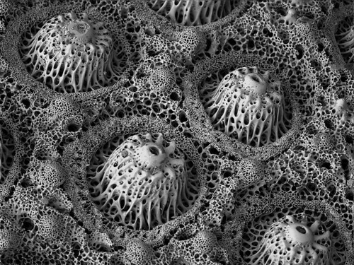
But what happens we direct an SEM at animals that are ALREADY kind of weird? The striking beauty of sea urchins is revelaed!
The sea urchin skeleton. Also known as a
TEST. One of my many regularly repeated caveats- these are NOT shells. These are underlying skeletons which have a layer of skin which is typically removed to reveal the more aesthetic skeleton...
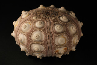
Here is an example from a cidaroid sea urchin.. Those round knobs or bosses? Those are where the spines articulate with the body...
 |
| Image by Gripspix |
Here's a nice macro shot of aforementioned "boss" (=the knob which connects with the spine).
 |
| Image by "Nervous System" |
But what happens when we focus a Scanning Electron Microscope (SEM) on these surfaces? (note that these images are a different species from the one above).
 |
| Image by Monkey.grip |
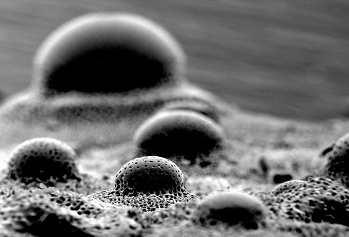
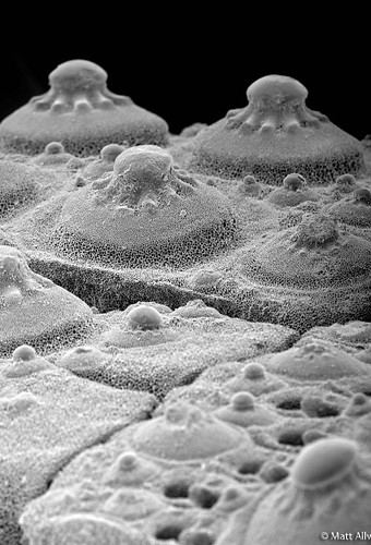
Here's another view looking down. Note how the skeleton is actually porous! Echinoderm skeletons aren't simply inorganic calcium carbonate, they are actually infused with tissue...
 |
| Image by gary.brake |
 |
| Image by Studio Jonas Coersmeier |
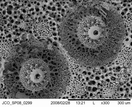
Another view from a different perspective...
 |
| Image by particlesixtyfour |
 |
| Image by monkey.grip |
Closer...
 |
| Image by particlesixtyfour |
..and closer still!
 |
| Image by particlesixtyfour |
right on top of it!
 |
| Image by particlesixtyfour |
But wait! What about the SPINES??? I'm not sure which species these are from.. but they give you a good idea about the fine topology that one might not realize is present
from simply looking at a spine with your eye...
 |
| Image by particlesixtyfour |
Closer!
 |
| Image by particlesixtyfour |
And finally... CLOSEST!!
 |
| Image by particlesixtyfour |
And just to cap off the whole odyssey through a sea urchin spine...
here is a cross section THROUGH a spine magnified 150X!!! The final three images below a
re from the Biology Dept. at the University of Dayton!
Here are the featured spines...
and together to show perspective...


4 comments:
These are beautiful! I read a neat paper by Dafni (1986) that talks about where test deposition occurs in the tissue, so some nice soft tissue work to go with the hard skeletons. Has anyone thought about why the tissue that deposits the skeleton is syncytial? Also, I posted a few SEMs from an irregular urchin a while back, if you want to see more SEM photos of urchin tests: http://bmscblog.wordpress.com/2012/09/08/getting-to-the-heart-of-urchin-spine-attachment/
Dear Chris:
A. Gaudi would be as enthralled as any of your echinoblog regulars/enthusiasts about the biomimicry possibilities and about the wondrous biophysiology that such SEM micrographs begin to reveal. Thank you much. FS
These look a lot like three-dimensional fractal images:
http://www.subblue.com/projects/mandelbulb
Wow! thanks Emily!
Post a Comment