Working late alone in the lab, going through a dish of marine inverts collected from a random sample in the Pacific Ocean and you find...something.
Sometimes someone is around to share it with, sometimes not. What does THAT feel like? This video isn't mine but it gives me the same feeling of discovery.
I just found this and I don't know what it is. THAT's saying something..
Enjoy!
Showing posts with label larvae. Show all posts
Showing posts with label larvae. Show all posts
Saturday, February 16, 2013


Tuesday, September 25, 2012
Fantastic pics of larvae and baby sea urchins!
Today..some great images of larval sea urchins from 2 researcher/photographers!
First some great video and images from Bruno C. Vellutini from Brazil and currently at the Sars International Centre for Marine Molecular Biology
Bruno is also an exceptional photographer and has been kind enough to allow his images to be shared with us.. Here are samples of his excellent work, much of which are of larval forms-that is the small swimming "babies" that form and settle out into the five-part adults that we are more familiar with..
Here is a quick video that shows the life cyle in a "sea biscuit" aka a sea urchin called Clypeaster subdepressus. Basically, egg and sperm meet, form free swimming bilateral larvae which then settles out on the sea bottom and grows up into a pentameral adult!
A Sea Biscuit's Life from Bruno Vellutini on Vimeo.
Larvae are very tiny.. and are on the order of about a 1.0 mm in length at best.
Here's some striking images from different points during the cycle...
The SKELETON of a larval Clypeaster urchin! Under polarized light..


Pluteus larva showing its left side

Still with 4 arms..

A neat color image showing both sides

A sea biscuit juvenile

A pic of a young sand dollar..

Here are some pics of various VERY tiny..baby urchins!
From Juan Camilo Jaramillo -a researcher at the Universidad Jorge Tadeo Lozano in Columbia! (on Flickr)
A larval urchin showing a spectacular glow!

A 1.0 mm Diadema (long black spined urchin when adult)

A small, newly settled unidentified urchin


NEXT WEEK! I promise some solid Echinoblogging!
First some great video and images from Bruno C. Vellutini from Brazil and currently at the Sars International Centre for Marine Molecular Biology
Bruno is also an exceptional photographer and has been kind enough to allow his images to be shared with us.. Here are samples of his excellent work, much of which are of larval forms-that is the small swimming "babies" that form and settle out into the five-part adults that we are more familiar with..
Here is a quick video that shows the life cyle in a "sea biscuit" aka a sea urchin called Clypeaster subdepressus. Basically, egg and sperm meet, form free swimming bilateral larvae which then settles out on the sea bottom and grows up into a pentameral adult!
A Sea Biscuit's Life from Bruno Vellutini on Vimeo.
Larvae are very tiny.. and are on the order of about a 1.0 mm in length at best.
Here's some striking images from different points during the cycle...
The SKELETON of a larval Clypeaster urchin! Under polarized light..
Pluteus larva showing its left side
Still with 4 arms..
A neat color image showing both sides
A sea biscuit juvenile
A pic of a young sand dollar..
Here are some pics of various VERY tiny..baby urchins!
From Juan Camilo Jaramillo -a researcher at the Universidad Jorge Tadeo Lozano in Columbia! (on Flickr)
A larval urchin showing a spectacular glow!
A 1.0 mm Diadema (long black spined urchin when adult)
A small, newly settled unidentified urchin
NEXT WEEK! I promise some solid Echinoblogging!
Thursday, August 26, 2010
Oil Vs. Echinoderms-the Video version!
So, here is the video version from Ocean Planet of my Echinoblog post on oil affecting echinoderms and the related Ocean Portal post on the same topic!! This usually lives here (on Ocean Portal).....Enjoy!
Tuesday, August 3, 2010
Cloning As Sand Dollar DEFENSE! Why run & hide when you can divide?
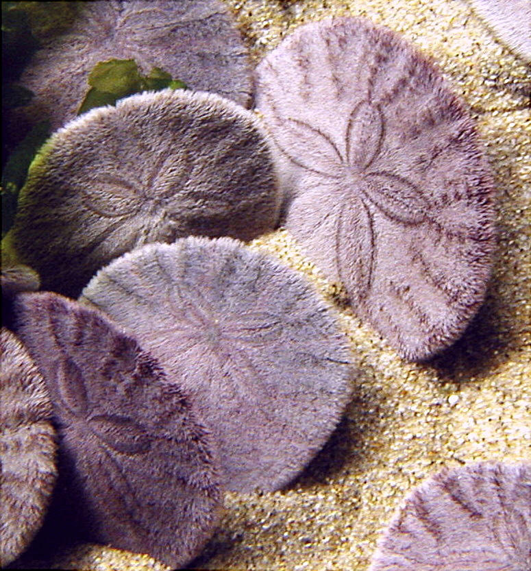 Today, a little somethin' somethin' about a familiar animal..but not quite the way, you're used to thinking about it.. Today's post is based on a new, interesting paper by Dawn Vaughn who is one of the many smart people at Friday Harbor Laboratories! This paper was in issue 157 of Marine Biology of this year (click here!).
Our subject is the familiar sea urchin-the Pacific Sand Dollar, Dendraster excentricus! Here is a good intro page. You can see pictures of the animal alive above..but here is how most people are accustomed to discovering it..
 Dendraster is a sea urchin-admittedly a very WEIRD sea urchin. and like all sea urchins (and indeed all echinoderms), the adult forms grow from a small larval stage that is so tiny, this asterisk * is probably slightly larger.
 A sea urchin larvae is called a pluteus (plural-plutei)..specifically an echinopluteus (for sea urchins).
 (Image borrowed from the Cephalopodiatrist!)
This larval stage floats in the sea before settling down and makes up part of the plankton. So, it was discovered some years ago, that these larvae can be in the water column for quite a LONG time. Months, possibly years. This can be important in the development of the adult..as we'll see..
These can often be food for other creatures that live in the ecosystem at about the same time.. Often, the larvae that float become FOOD for other organisms, such as crustaceans and fish.
Recently though, larval life has been thought of as kind of an added "secret" life and are capable of doing much more then simply floating around, waiting to be eaten or becoming adults.
 |
| (Image borrowed from the Cephalopodiatrist!) |
It turns out that SOME echinoderm larvae, sea stars and sea urchins (probably others) can actually CLONE themselves. That is to say, they can "reproduce" asexually when they are larvae. (Adult sand dollars cannot divide in half and clone themselves in the same way..)
But WHAT would the evolutionary significance of this action/behavior be??? How does it help the animal survive?
In some studies, echinoderm larvae clone themselves when times are good. Food is prime and temperature is ideal.
But what happens we investigate cloning in the context of hostile or high-risk environments???
There are obvious advantages to being able to clone oneself in the face of danger from predators, such as safety in numbers and so on.
To investigate these questions Vaughn set up an interesting experiment.
They cultured the plutei from Dendraster excentricus and cleverly created an experiment that simulated the presence of different, local fish species in the environment!
 How did they do that? They isolated and concentrated the fish MUCUS from 2 different species-the stickleback (above) and Dover sole (Microstomus pacificus).
One of the groups was a "control" group-i.e., fish were not encouraged to attack, whereas the other group had mucus introduced as the "hostile" environmental factor.
Vaughn identified the presence of cloned Dendraster based on the the increased density of larvae (these were in a closed area after all) and the presence of larvae showing physical traces of budding. So, indirect and direct ways of determining the presence of "Uncloned" versus "Cloned"
Here's what they got...
 (Figure 2 from Vaughn, 2010)
Vaughn found that all of the larvae left in the control (without fish mucus to "threaten" them) were much fewer and much LARGER. Mucus-treated larvae INCREASED in numbers and DECREASED in SIZE over the duration of the experiment.
Figure 2 above shows a nice scale comparison of plutei from the Uncloned versus Cloned subjects.
Take-Away Message #1: When threatened, Dendraster larvae are apparently ENCOURAGED to clone themselves more! Cloned Dendraster are smaller.
Vaughn also performed trials where they looked at direct effect that fish attacks would have on the larvae.
On the graph below, the black bars represent UNCLONED larvae eaten whereas the grey bars represent CLONED larvae eaten.  (Figure 5 from Vaughn, 2010)
The UNCLONED (and LARGER) larvae (in black) suffered substantially GREATER losses to attacking fish then those CLONED (and SMALLER) larvae (in grey).
So, the rather strongly suggests that being able to clone oneself (thus creating MORE and SMALLER) individuals is an adaptive defense. (I suppose that's kind of a bummer if you're one of those larger larvae that get eaten though!)
Bear in mind the time scales here also... The asexual cloning doesn't work on an individual-individual instant scale..i.e., fish approaches and larvae splits. But when the fish leaves a mucous presence in the area..it presumably sets all of the larvae on alert to begin cloning themselves.
This can have a potentially important evolutionary and ecological effect. The presence of more larvae suggests that more fish could be present. Is escalation present? What is the effect of having these cloned larvae on the variation and evolution of the adults?

Friday, July 16, 2010
Pycnopodia Juvenile Stage Has Arrived!!
July 16 UPDATE!!! by Allison Gong:
What does Pycnopodia look like before it starts growing into an "adult"???
 My colleague Dr. Allison Gong at the UC Santa Cruz Long Marine Laboratory recently had the fortune to observe the spawning and early larval development of Pycnopodia helianthoides-the sunflower star one of my favorite animals!
 (This and all pictures courtesy of Allison Gong, UCSC)
Info and the comments below are graciously provided by her (seen above in her natural habitat)!
Apparently the gametes collected were produced by these hefty beasts on display at the Seymour Center. There are four in the aquarium. Each one may be either the mother or the father of these larvae.
(This and all pictures courtesy of Allison Gong, UCSC)
Allison says: "This is a 3-day-old early dipleurula larva. It is basically a ciliated blob with an invagination in the flattened posterior end. The internal tube and knob structures are the developing larval gut."
 (This and all pictures courtesy of Allison Gong, UCSC)
 (This and all pictures courtesy of Allison Gong, UCSC)
 (This and all pictures courtesy of Allison Gong, UCSC)
 (This and all pictures courtesy of Allison Gong, UCSC)
UPDATE Ladies and Gentlemen, we have BIPINNARIA!!! (an intermediate larval stage of starfish)
 Sez Allison: "These guys sure don't develop as quickly as urchins! At 14 days, the larva has reach a stage called the bipinnaria. It's sort of a more elaborate version of the dipleurula. In this ventral view, you can see the stomach quite clearly as the darkish ovoid shape in the bottom part."
 "Here's a left-side view of a different larva at the same stage. These guys are really transparent, and it's hard to get a feel for their three-dimensional structure because the camera focuses on a single plane."
 "These guys happened to swim under the objective lens at the same time, so I snapped a shot of them. In this photo you're getting both a ventral and a dorso-lateral view."
 10 June 2009-Allison Sez: These larvae are growing so slowly! I'm used to things happening more quickly. Oh well. This is a ventral (front) view of a 22-day-old bipinnaria larva. The internal ovoid structure in the lower half is the stomach, with dark food cell visible inside. Here's a trick to "seeing" the 3-dimensional structure in a 2-d photo: Imagine a capital letter 'c' and rotate it 90 degrees along its vertical axis. You're now looking into the "opening" of the 'c', right? That's exactly the view of this larva. The roughly triangular object on the top and the squarish object on the bottom correspond to the ends of the 'c'. Confusing, isn't it?
 Okay, here's a nice lateral view. The gut is nicely visible in this picture. You can see the elongated esophagus and the sphincter where it meets the stomach. You are looking at the larva's left side, with its anterior end up and its posterior end down.
 Bipinnaria larvae reaches the 29 day stage!
Sez Allison: We had a near miss late last week and lost about half of the larvae. I think maybe they didn't like the food we were giving them. We've altered the diet and the remaining larvae seem happy, although some of them may be arrested at an early stage of development.
This individual was the most advanced of all the larvae I observed today. At 29 days it is starting to develop little nubbins that may grow into long brachiolar arms--at least, I hope they do!--and measures 850 microns in length.
 Sez Allison: This larva, at the ripe old age of 49 days, has reached the brachiolaria stage. It measures a whopping 1.5 mm long! The gut is a golden color because we've been feeding them a mixture of green algae and diatoms.
The simple band in the earlier stages has been elaborated into pointy little nubbins called brachiolar arms. In other species, such as Pisaster ochraceus, the arms get really long; I don't know how long they'll get in these Pycnopodia larvae. We'll have to wait and see.
 Sez Allison: Whoa. Huge progress made in the last week! The larvae are 55 days old now and have *finally* started growing the long arms we've been expecting for weeks. I shot this photo through my Wild dissecting scope to get the cool effect of a dark background. Couldn't entirely get rid of the unwanted glare, though. Oh well.
 Sez Allison: Here's a closer shot of the same larva. The brownish structure in the bottom of the larva is its stomach.
 From Allison: Isn't this gorgeous? At 66 days (7 weeks and counting) the larvae have reached the advanced bracholaria stage. The arms are longer and the larvae wave them as they swim around. These guys are still feeding, although they haven't grown much. It looks like they max out at about 1.5 mm in length. This particular larva may not be entirely competent (i.e., ready to metamorphose) yet, but it's getting close.
 SETTLEMENT!!!
From Allison: Ta-dah! One larva has begun the process of metamorphosis. It has attached itself to a small piece of mussel shell with some suckers on the anterior end. The round structure you see at the top of the animal is the juvenile rudiment, or the earliest stage of the juvenile body, containing the water vascular system's first 5 tube feet. You can see that the brachiolar arms are still there. At this point the critter can no longer feed, as it completely re-arranges its entire body, and survives on energy reserves it put away as a feeding larva.
I never get tired of watching this kind of metamorphosis, in stars or urchins. It's the coolest thing out there. In a matter of a few days the animal transforms from a bilateral swimming creature to a pentaradial crawling beast, with a full scale re-arrangement of its external and internal anatomy. Larval parts will be resorbed or discarded, and new juvenile structures will be formed.
Amazing, isn't it? From Allison: A star is born! The little guy we've been monitoring, seen here in side view, has completed metamorphosis, and as far as I can tell is doing fine. All of the larval body has been resorbed now, and the critter is a little round disc with tube feet and spines. It's not very active right now but I think that's because it's recovering from the trauma of metamorphosis.
 From Allison: I tried to get a better view of the aboral (i.e., top) surface but immediately lost contrast because the baby star is almost the same color as the bit of mussel shell it's sitting on. At least in this view you can see that it's radial now. Quite a change from the bilateral larva it was a week ago, isn't it?
I think these new juveniles will fast for a number of weeks, living on energy reserves they packed away while they were feeding larvae. It remains to be seen whether or not we can figure out what to feed these tiny guys, but we'll try to keep them going and will hopefully be able to document how all of the arms develop. Remember, these stars have ~20 arms as adults, although they start out with the requisite echinoderm 5, and we're interested in seeing if there's a pattern to how all the arms form.
 So, a late addendum... have you ever wondered what Pycnopodia looks like when they are just wee babies?? Here ya' go..
 According to Allison, this is what they look like 23 days past settlement. They have FIVE rays just like ANY other SEA STAR. Neat, eh??
From here, they grow onto the enormous, multi-rayed forms that we know and love! I suspect that takes AT LEAST several months to years.... so, leave em' alone when you find em as adults! It probably takes a LONG time for them to reach even a 6 inch diameter!
What does Pycnopodia look like before it starts growing into an "adult"???

 (This and all pictures courtesy of Allison Gong, UCSC)
Info and the comments below are graciously provided by her (seen above in her natural habitat)!
Apparently the gametes collected were produced by these hefty beasts on display at the Seymour Center. There are four in the aquarium. Each one may be either the mother or the father of these larvae.
(This and all pictures courtesy of Allison Gong, UCSC)Allison says: "This is a 3-day-old early dipleurula larva. It is basically a ciliated blob with an invagination in the flattened posterior end. The internal tube and knob structures are the developing larval gut."
 (This and all pictures courtesy of Allison Gong, UCSC)
Allison says: "This photo is essentially the same thing, photographed under dark-field conditions for a rather cool effect."
 (This and all pictures courtesy of Allison Gong, UCSC)
A neat black and white shot...
 (This and all pictures courtesy of Allison Gong, UCSC)
Two More!!
 (This and all pictures courtesy of Allison Gong, UCSC)
UPDATE Ladies and Gentlemen, we have BIPINNARIA!!! (an intermediate larval stage of starfish)
 Sez Allison: "These guys sure don't develop as quickly as urchins! At 14 days, the larva has reach a stage called the bipinnaria. It's sort of a more elaborate version of the dipleurula. In this ventral view, you can see the stomach quite clearly as the darkish ovoid shape in the bottom part."
 "Here's a left-side view of a different larva at the same stage. These guys are really transparent, and it's hard to get a feel for their three-dimensional structure because the camera focuses on a single plane."
 "These guys happened to swim under the objective lens at the same time, so I snapped a shot of them. In this photo you're getting both a ventral and a dorso-lateral view."
 10 June 2009-Allison Sez: These larvae are growing so slowly! I'm used to things happening more quickly. Oh well. This is a ventral (front) view of a 22-day-old bipinnaria larva. The internal ovoid structure in the lower half is the stomach, with dark food cell visible inside. Here's a trick to "seeing" the 3-dimensional structure in a 2-d photo: Imagine a capital letter 'c' and rotate it 90 degrees along its vertical axis. You're now looking into the "opening" of the 'c', right? That's exactly the view of this larva. The roughly triangular object on the top and the squarish object on the bottom correspond to the ends of the 'c'. Confusing, isn't it?
 Okay, here's a nice lateral view. The gut is nicely visible in this picture. You can see the elongated esophagus and the sphincter where it meets the stomach. You are looking at the larva's left side, with its anterior end up and its posterior end down.
 Bipinnaria larvae reaches the 29 day stage!
Sez Allison: We had a near miss late last week and lost about half of the larvae. I think maybe they didn't like the food we were giving them. We've altered the diet and the remaining larvae seem happy, although some of them may be arrested at an early stage of development.
This individual was the most advanced of all the larvae I observed today. At 29 days it is starting to develop little nubbins that may grow into long brachiolar arms--at least, I hope they do!--and measures 850 microns in length.
 Sez Allison: This larva, at the ripe old age of 49 days, has reached the brachiolaria stage. It measures a whopping 1.5 mm long! The gut is a golden color because we've been feeding them a mixture of green algae and diatoms.
The simple band in the earlier stages has been elaborated into pointy little nubbins called brachiolar arms. In other species, such as Pisaster ochraceus, the arms get really long; I don't know how long they'll get in these Pycnopodia larvae. We'll have to wait and see.
 Sez Allison: Whoa. Huge progress made in the last week! The larvae are 55 days old now and have *finally* started growing the long arms we've been expecting for weeks. I shot this photo through my Wild dissecting scope to get the cool effect of a dark background. Couldn't entirely get rid of the unwanted glare, though. Oh well.
 Sez Allison: Here's a closer shot of the same larva. The brownish structure in the bottom of the larva is its stomach.
 From Allison: Isn't this gorgeous? At 66 days (7 weeks and counting) the larvae have reached the advanced bracholaria stage. The arms are longer and the larvae wave them as they swim around. These guys are still feeding, although they haven't grown much. It looks like they max out at about 1.5 mm in length. This particular larva may not be entirely competent (i.e., ready to metamorphose) yet, but it's getting close.
 SETTLEMENT!!!
From Allison: Ta-dah! One larva has begun the process of metamorphosis. It has attached itself to a small piece of mussel shell with some suckers on the anterior end. The round structure you see at the top of the animal is the juvenile rudiment, or the earliest stage of the juvenile body, containing the water vascular system's first 5 tube feet. You can see that the brachiolar arms are still there. At this point the critter can no longer feed, as it completely re-arranges its entire body, and survives on energy reserves it put away as a feeding larva.
I never get tired of watching this kind of metamorphosis, in stars or urchins. It's the coolest thing out there. In a matter of a few days the animal transforms from a bilateral swimming creature to a pentaradial crawling beast, with a full scale re-arrangement of its external and internal anatomy. Larval parts will be resorbed or discarded, and new juvenile structures will be formed.
Amazing, isn't it?
 From Allison: A star is born! The little guy we've been monitoring, seen here in side view, has completed metamorphosis, and as far as I can tell is doing fine. All of the larval body has been resorbed now, and the critter is a little round disc with tube feet and spines. It's not very active right now but I think that's because it's recovering from the trauma of metamorphosis.
 From Allison: I tried to get a better view of the aboral (i.e., top) surface but immediately lost contrast because the baby star is almost the same color as the bit of mussel shell it's sitting on. At least in this view you can see that it's radial now. Quite a change from the bilateral larva it was a week ago, isn't it?
I think these new juveniles will fast for a number of weeks, living on energy reserves they packed away while they were feeding larvae. It remains to be seen whether or not we can figure out what to feed these tiny guys, but we'll try to keep them going and will hopefully be able to document how all of the arms develop. Remember, these stars have ~20 arms as adults, although they start out with the requisite echinoderm 5, and we're interested in seeing if there's a pattern to how all the arms form.
 So, a late addendum... have you ever wondered what Pycnopodia looks like when they are just wee babies?? Here ya' go..
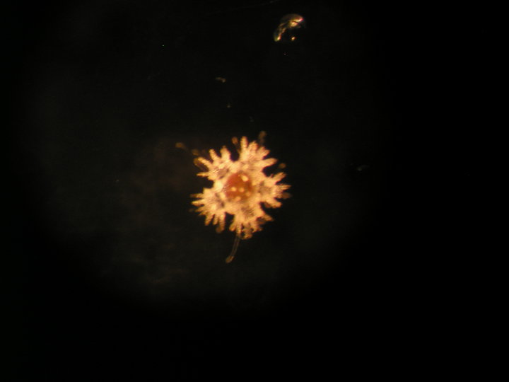 According to Allison, this is what they look like 23 days past settlement. They have FIVE rays just like ANY other SEA STAR. Neat, eh??
From here, they grow onto the enormous, multi-rayed forms that we know and love! I suspect that takes AT LEAST several months to years.... so, leave em' alone when you find em as adults! It probably takes a LONG time for them to reach even a 6 inch diameter!
Monday, November 9, 2009
Tosia Times TWO! Discovering NEW CRYPTIC species of Australian Biscuit Stars!
 |
| Image courtesy of Kate M. Naughton! |
Today's blog comes from my colleagues Kate Naughton and Tim O'Hara at the Museum Victoria in Melbourne, Australia! who have JUST published a paper describing a NEW species of the familiar Australian biscuit starfish, Tosia in Invertebrate Systematics!
 (one is Kate and one is Tim-can you guess which one?)
Tosia, as I define it, is found ONLY in Australia, where it is quite abundant and relatively well-known to the people who study marine biology thereabouts. One species in particular, Tosia australis, occurs widely throughout the temperate southern part of Australia.
 (Image courtesy of Kate M. Naughton!)
It has led to the identification of what's called the Tosia australis species complex!! That's what happens when you have a lot of closely-related populations that all may or may not be different species.
These populations usually show tenuous morphological differences between different "species" in the related network which are typically spread out over a relatively large geographic region. The North Pacific 6-rayed starfish Leptasterias, which I've written about here, is also a species complex.
Something NEW! Brooding Discovered!
So, for an animal that occurs so close to shore, surprisingly little was known about it. In the 90s it was discovered that Tosia australis was a SPECIAL kind of starfish!! It showed an unusual reproductive behavior: BROODING juveniles! That is to say, its offspring live on the adults.
Not too unusual in vertebrates, but VERY unusual in starfish!
Also, Tosia australis has been known since 1840-so it took over 150 years for them to discover that it brooded juveniles!! So, this led to some close observation.

 (Image courtesy of Kate M. Naughton!)
So, the investigative talents of Ms. Naughton and Dr. O'Hara were applied towards the question!
Did the difference in larvae mode also translate into further biological differences??
During a museum visit to Melbourne, I was talking shop with Kate and Tim. I was pretty sure that all the morphological variation in the species complex would translate into regional or environmental differentiation VS. Tim who believed that they would split out by larval type.
Larvae in several other species is highly variable but often did not translate into phylogenetic differences..so I figured it would probably do the same here.
And in fact, I was SO confident I was right, that I wagered a gentlemen's bet with Dr. O'Hara that I would be right!
So-I said: "Go get the DNA and prove me wrong!"
 Oops! Guess I just lost 10 bucks!
The final analysis following a survey of many specimens from Victoria, Tasmania, and South Australia supported three different species, including something NEW that hadn't been observed before!!!
 (Phylogenetic Tree inferred from COI and 16S RNA-and redrawn here by the Echinoblog Art Department!)
Several specimens from Tasmania and Victoria were supported on a separate clade as a NEW SPECIES. Kate came to call it Tosia neossia. The species epithet neossia is Greek for "nest" in reference to this species' brooding habit.
And here it is!
 (Image courtesy of Kate M. Naughton!)
T. neossia is very similar to the well-known T. australis but, aside from a bunch of external morphological features, one important feature sets them apart....
(Image courtesy of Kate M. Naughton!)
T. neossia is very similar to the well-known T. australis but, aside from a bunch of external morphological features, one important feature sets them apart....
The difference is in the Larvae (i.e., juvenile starfish)!
And here it is!
 (Image courtesy of Kate M. Naughton!)
The difference is in the Larvae (i.e., juvenile starfish)!
One of the fundamental differences here is that of the difference in larvae between Tosia australis and T. neossia!
(Image courtesy of Kate M. Naughton!)They are emitted from the mother and crawl along the bottom using little lobes to get around until they fully develop into larval adults. These little BOTTOM larvae (shown here as early stage larvae) can be seen here (courtesy of Kate Naughton!)
 (Image courtesy of Kate M. Naughton!)
Here is where the larvae begin to change into what Kate calls the "tripod phase". These utilize a large lobe that helps move the larvae around before it begins to attach someplace on the ground after developing to the right stage....
 (Image courtesy of Kate M. Naughton!)
This little fellow will develop into what will eventually become
 (Image courtesy of Kate M. Naughton!)
....the adult!
This is in direct CONTRAST to Tosia australis which has SWIMMING juveniles that emerge from the aboral surface (via gonopores on the top surface). In contrast to the larval juveniles of T. neossia, T. australis has eggs that are postively buoyant and are covered with small CILIA!! That is, they have little beating hair-like threads that can keep them swimming before they settle out somewhere.Summary Time!
In addition to the DNA and morphological evidence (you can go to the paper for details) the larval story goes like this:
Tosia neossia! Gonopores open on ORAL surface (i.e., DOWN) where the juveniles sink to the bottom. They are bottom-living and non-ciliated until they grow into juvenile adults!
Tosia australis! Gonopores open on ABORAL surface (i.e., UP) where the juveniles float and can sustain a swimming stage until they settle to the bottom and grow into juvenile adults
TWO species. VERY similar looking! but with DISTINCT juveniles growing into adults that are hiding right in plain sight!
Best 10 bucks I ever lost.
Thanks to Kate Naughton for helping me with images!
Subscribe to:
Posts (Atom)